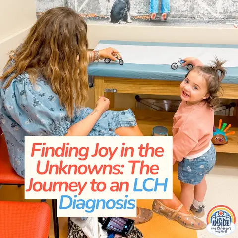
Navigating an LCH Diagnosis in Toddlers: What Parents Need to Know
Navigating an LCH diagnosis in toddlers can feel like an isolating and terrifying journey for medical parents. This guide breaks down the early warning signs, the intricacies of rare bone lesion identification, and what families can expect during the diagnostic and staging process for Langerhans Cell Histiocytosis.
Key Takeaways
- LCH (Langerhans Cell Histiocytosis) in toddlers often presents subtly, starting with unexplained bone pain or tenderness during diaper changes.
- Initial X-rays can sometimes misdiagnose LCH lesions as bone cysts or hairline fractures, making specialized pediatric orthopedic review critical.
- A full-body skeletal survey and targeted MRIs help determine whether LCH is single-system or multi-system, guiding treatment options.
- Parents often face overwhelming waits for biopsy results and staging, but managing the emotional spiral is just as important as medical research.
- Advanced pediatric centers like CHOP offer specialized pathways, sometimes allowing eligible toddlers to start with oral chemotherapy rather than port placement.
Recognizing the Early Signs of LCH in Toddlers
When a baby or toddler begins experiencing unexplained pain, parents naturally look for common culprits: growing pains, a minor tumble off the couch, or a muscle strain. However, for parents navigating an LCH diagnosis in toddlers, the presentation is often distinct yet easy to misattribute. In many cases, the first indicators are behavioral shifts during routine caregiving tasks.
Parents might notice their previously happy baby crying out inconsolably during diaper changes, getting dressed, or being lifted from a high chair. When physical tenderness centers around weight-bearing bones like the femur or hips without any visible bruising or swelling, it serves as a critical red flag. As toddlers grow and begin to pull themselves up or walk, parents may observe a sudden regression, such as an unusual limp, a reluctance to climb stairs, or difficulty maintaining balance.
The Diagnostic Maze: From Bone Cysts to LCH Confirmation
The path to an accurate LCH diagnosis in toddlers is rarely straightforward. Because Langerhans Cell Histiocytosis is exceptionally rare, primary care pediatricians and local imaging centers may initially interpret bone lesions as something much more common, such as a simple bone cyst accompanied by a hairline fracture.
Getting a second opinion or having imaging reviewed by specialized pediatric orthopedic oncologists can completely alter the trajectory of care. Without expert eyes reviewing the films, a child might be unnecessarily casted for a suspected fracture, delaying the true diagnosis. Once a physician suspects a lesion rather than a cyst, the diagnostic protocol moves quickly into specialized territory.
The Role of Biopsies and Full-Body Scans
Confirming an LCH diagnosis requires a definitive tissue sample, typically gathered through a minimally invasive needle biopsy performed under anesthesia. Once the pathology report confirms the presence of abnormal white blood cells, the medical team must determine the extent of the disease.
This staging process involves comprehensive imaging, including:
- Full-body skeletal surveys (head-to-toe X-rays) to check for other bone involvement.
- Targeted MRIs of high-risk areas like the hips, femur, and spine.
- Organ imaging to ensure the liver, spleen, lungs, and pituitary gland are unaffected.
Discovering that the disease is confined strictly to the bone can bring immense relief, though ongoing monitoring remains essential because lesions can occasionally spread over time, requiring a shift from watchful waiting to active treatment.
Coping With the Emotional Whiplash of Rare Disease Parenting
Receiving a call about a rare disease diagnosis on a Friday afternoon triggers an immediate, overwhelming sense of panic. For parents staring down the acronym LCH for the very first time, the natural instinct is to turn to the internet for answers. Unfortunately, search engine results often highlight worst-case scenarios, fueling a relentless emotional spiral.
Medical parents quickly learn that pacing themselves is a survival skill. You do not need to master every medical journal article about white blood cell disorders in twenty-four hours. Building a trusted relationship with a dedicated pediatric oncology team and connecting with other rare disease families can transform an isolating experience into one grounded in community and hope.
Moving Forward With Hope and Connection
While an LCH diagnosis in toddlers alters family life overnight, it does not define a child's entire future. With specialized care, supportive medical teams, and a growing network of rare disease advocates, families can navigate the unknowns with resilience. To hear more about one mother's real-time experience balancing hospital visits, family life, and finding joy in the small moments, Listen to the full episode. Tune in to gain deeper insights and compassionate encouragement for the pediatric healthcare journey.
Frequently Asked Questions
What are the early warning signs of LCH in toddlers?
Early signs of Langerhans Cell Histiocytosis (LCH) in toddlers often include localized bone pain, tenderness when dressing or changing diapers, unexplained limping, or developmental regression in mobility like trouble walking up steps.
Why is LCH frequently misdiagnosed initially?
LCH lesions in bones can mimic common pediatric issues like benign bone cysts or hairline fractures on standard X-rays. Specialized evaluation by pediatric orthopedic oncology experts is often required to correctly identify the lesions.
Is LCH considered a form of childhood cancer?
LCH is technically a rare inflammatory myeloid neoplasm characterized by the overproduction of white blood cells, but it is typically managed and treated through pediatric oncology protocols.
What diagnostic tests are used after an LCH suspicion?
Once LCH is suspected, doctors typically order a needle biopsy for confirmation, followed by a full-body skeletal survey, MRIs of affected joints and bones, and organ imaging to determine if the disease is single-system or multi-system.